Article -> Article Details
| Title | What Imaging Is Used Before Breast Augmentation? |
|---|---|
| Category | Fitness Health --> Health Articles |
| Meta Keywords | breast augmentation dubai price |
| Owner | Ahmad |
| Description | |
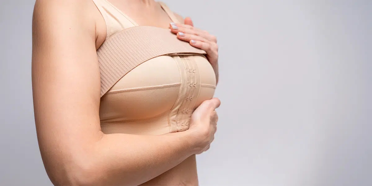
| What Imaging Is Used Before Breast Augmentation? is an important consideration for patients planning cosmetic breast procedures, especially when aiming for safe and predictable outcomes. In modern aesthetic practice, careful imaging helps evaluate breast tissue, chest anatomy, and implant planning. Many patients searching for cost of breast augmentation dubai also want to understand what diagnostic steps are involved before surgery, particularly in advanced clinics such as Dynamic Life Clinic where pre-operative evaluation is prioritized to support clinical accuracy and patient safety. Importance of Pre-Operative Imaging in Breast AugmentationPre-operative imaging plays a key role in ensuring that breast augmentation is performed with precision and safety. It allows specialists to understand internal breast structures, detect any underlying conditions, and choose the most suitable surgical approach. Imaging is not just about visualization; it is about planning. Each patient has unique anatomy, and imaging helps map:
This detailed assessment reduces uncertainty during surgery and supports more predictable aesthetic outcomes.
Common Imaging Techniques Used Before Breast AugmentationSeveral imaging tools may be used depending on age, medical history, and clinical findings. Each technique serves a different purpose in pre-surgical evaluation. MammographyMammography is a specialized X-ray of the breast used primarily to screen for abnormalities before surgery. Key purposes include:
It is commonly recommended for patients above a certain age or those with risk factors. Breast UltrasoundUltrasound is widely used in pre-operative breast assessment due to its ability to provide real-time imaging without radiation. It helps to:
This imaging method is often the first step in evaluating breast anatomy. Magnetic Resonance Imaging (MRI)MRI provides highly detailed imaging and is used in more complex or high-risk cases. It is particularly useful for:
MRI offers a comprehensive view of both soft tissue and surrounding structures, making it valuable in advanced surgical planning. Role of 3D Imaging and Digital Breast AnalysisIn modern cosmetic practice, 3D imaging systems are increasingly used to simulate surgical outcomes. These tools do not replace medical imaging but complement it by providing visual planning support. Benefits include:
This technology helps align patient expectations with realistic surgical possibilities. Safety Assessment Through ImagingImaging before breast augmentation is closely linked to patient safety. It ensures that any hidden medical conditions are identified before surgery begins. This step significantly reduces surgical risks and supports informed decision-making. Safety-focused imaging helps in:
By integrating imaging into pre-operative planning, specialists can tailor procedures with greater precision. How Imaging Supports Surgical PlanningBreast augmentation is not a one-size-fits-all procedure. Imaging provides the anatomical roadmap needed to customize the surgical approach. It supports:
This planning stage ensures that surgical decisions are based on clinical data rather than visual estimation alone. What Patients Should Expect Before ImagingBefore undergoing imaging for breast augmentation planning, patients may be advised to follow certain guidelines depending on the type of scan. General expectations include:
These steps help ensure accurate imaging results and smoother evaluation. Integration of Imaging in Modern Aesthetic PracticeIn advanced cosmetic environments, imaging is considered an essential part of the pre-surgical workflow. It supports both medical accuracy and aesthetic precision, ensuring that the final outcome aligns with the patient’s body structure and expectations. Clinicians rely on imaging not only to plan surgery but also to build a complete understanding of each patient’s unique anatomy. This contributes to safer procedures and more refined results in breast augmentation cases. ConclusionPre-operative imaging is a fundamental step in breast augmentation planning, offering detailed insights into breast anatomy, tissue health, and structural symmetry. Techniques such as mammography, ultrasound, MRI, and 3D imaging work together to support safe and personalized surgical outcomes. By combining medical evaluation with advanced imaging tools, specialists can design procedures that align with both clinical requirements and aesthetic goals, ensuring a more predictable and well-structured surgical experience. | |